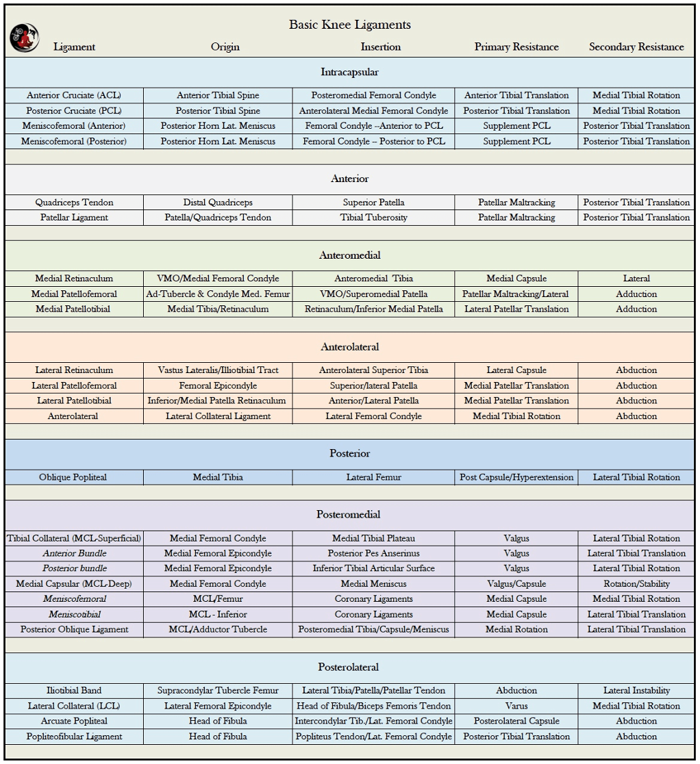
Muscles and Ligaments
Ligaments and Muscles that Cross the Knee Joint Ligaments
Knee ligaments provide resistance to internal and external forces to ensure optimal joint function, congruency and stability. These soft tissue structures are comprised of dense collagen fibers with predictable mechanical features as a function of shape, structure and organization.
The elasticity and tensile strength of each ligament is specific to its intended function.
Cruciate Ligaments
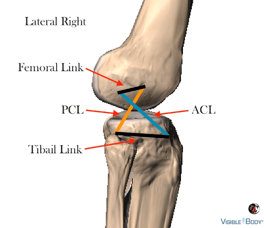
The cruciate ligaments restrain anterior and posterior translations of the tibia and femur essential to the glide component of tibiofemoral arthrokinematics. Barring these contributions the bones would simply roll off one another during flexion and extension.
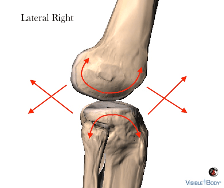
The orientation and function of the cruciates, tibia and femur can be compared to a four-bar linkage mechanism, though the ligamentous connections are not entirely rigid like the tibial and femoral links.
A four-bar mechanism is a closed chain linkage system that including four levers and hinges to control movement, improve function and stability. Examples include bicycle suspension, vice grips, oil well pumps, etc.
Glute contraction is transmitted to the femur, which acts as a driver to convert power into tension at the ACL and PCL, assisting femoral rotation on the tibia during extension.
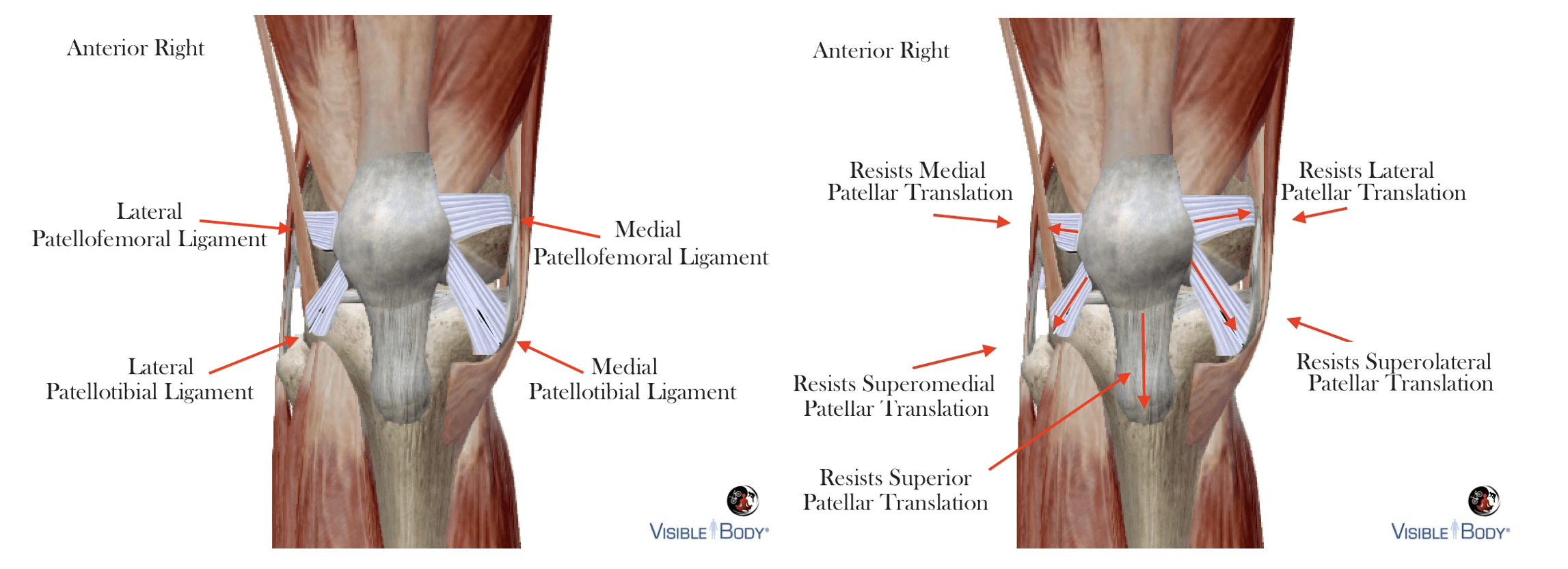
Anterior Ligaments
The patellar ligament links the quadriceps muscles to the tibia (via patella) for knee extension. Optimal positioning reduces compressive patellofemoral forces produced by the quadriceps during extension.
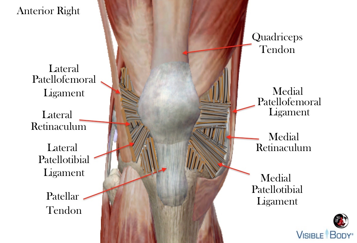
Patellofemoral and patellotibial ligaments work to keep the patella tracking properly the trochlear groove. The medial patellofemoral ligament provides the greatest resistance to lateral patellar displacement, though we cannot assume that a laterally offset patella is “maltracking” because some folks are just born that way;).
Posterior Ligaments
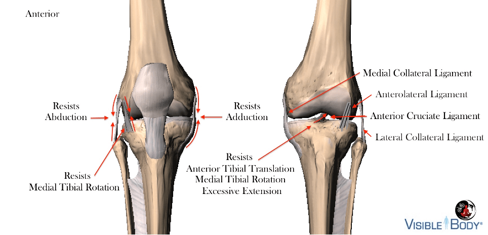
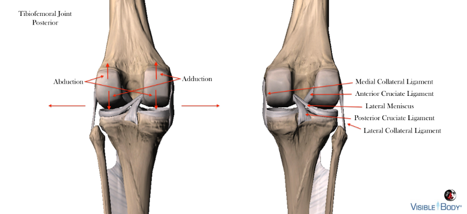
The collateral ligaments are recognized primarily in the context of abduction/adduction stability – resisting excessive varus/valgus knee angles. The terms medial collateral ligament/tibial collateral ligament and lateral collateral ligament/fibular collateral ligament are interchangeable, with the latter referring to the insertion points below the knee.
We are more concerned with the medial collateral ligament when working with cyclists, because it contains deep fibers that attach to the medial menisci and blends with the joint capsule. Adjusting a rider’s stance and foot correction to avoid excessive valgus knee/adductor moments is vital to capsular and meniscus integrity.
The anterior cruciate and anterolateral ligaments moderate medial tibia on femur rotation during extension. Excessive medial tibial rotation tends to accompany abnormal q-angles/genu valgum, pronation, ankle instability, etc., and responds positively to foot correction/orthosis assuming optimal pedaling base.
The minescofemoral ligament connects the posterior horn of the lateral meniscus to the lateral aspect of the medial femoral condyle. It splits into the anterior meniscofemoral ligament (ligament of Humprhrey) and posterior meniscofemoral ligament (ligament of Wrisberg) at the posterior cruciate ligament. These ligaments contribute to checking external tibial rotation, which typically occurs in a toe-out foot position typical of a wide pedaling base or morphology. We can discern between the two-conditions because the foot angle will straighten with reduced pedaling base (assuming sufficient float/cleat adjustment) when driven by fit and not anatomy.
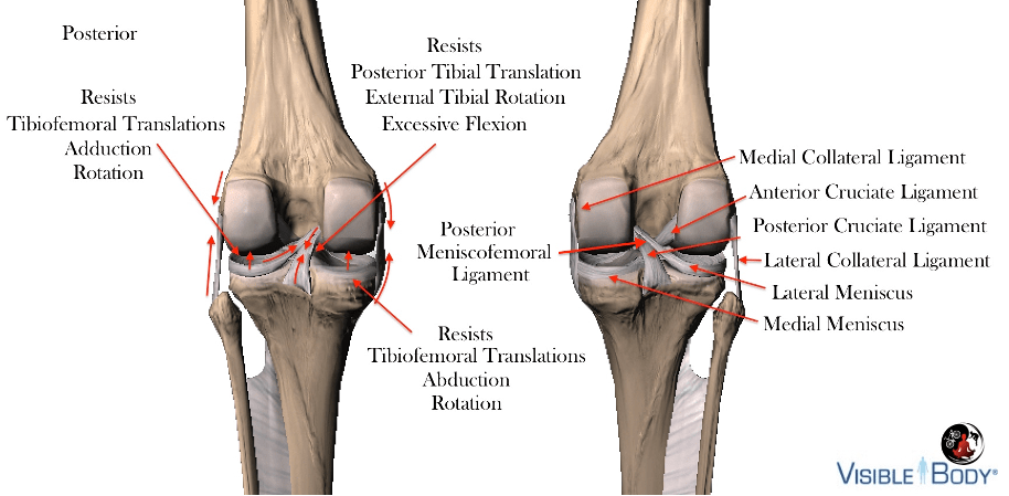
Contralateral menisci compression accompanies medial and lateral collateral ligament tension when coupled with excessive varus and valgus moments.
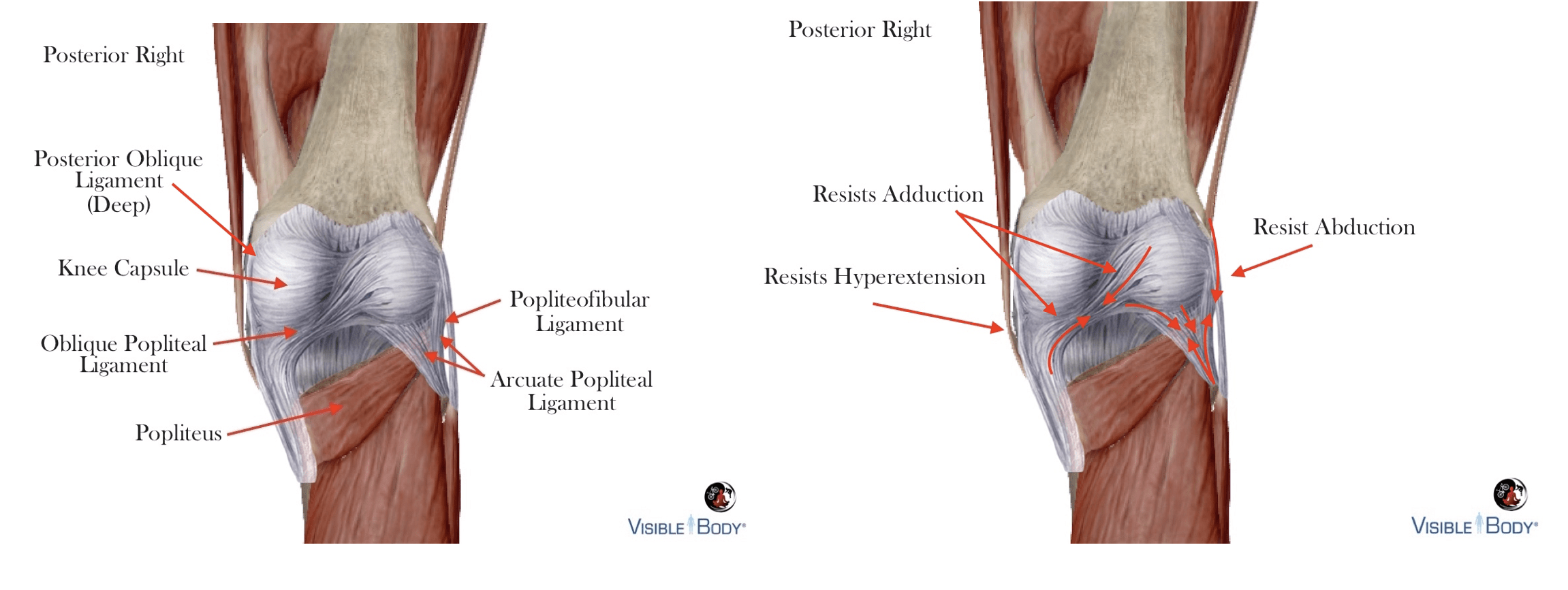
The posterior capsular and supporting ligaments resist angular and rotational forces at the knee joint with emphasis on resisting hyperextension and supplementing the fibular collateral ligament at posterolateral compartment
Muscles
Muscles that cross the knee produce flexion and extension with additional frontal and transverse plane motions. Appropriate pedaling base, rotation and foot corrections should promote primary function and balanced secondary movement patterns
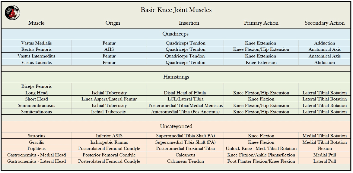
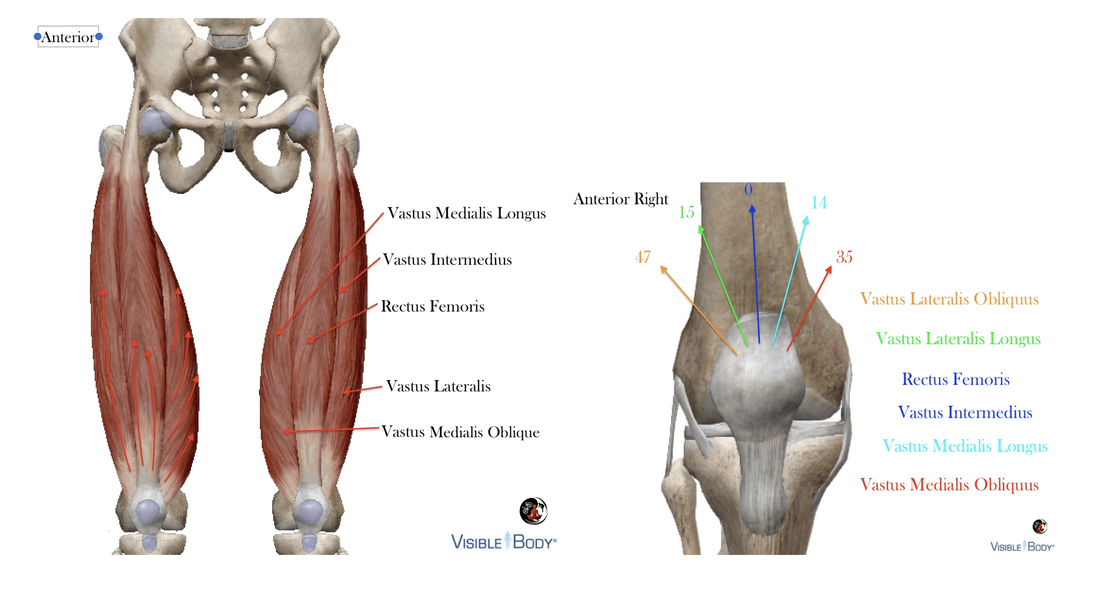
Anterior Knee Muscles
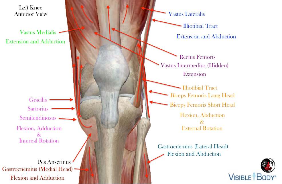
Each quadriceps muscle imposes a unique line of pull on the patella that is consistent with the direction of muscle fiber and orientation of the joint in the frontal plane. The fibers in muscles like the vastus medialis change direction from origin to insertion with increased mediolateral pull on the patella at quadriceps tendon.
A frontal plane medal or lateral shift in knee joint position alters the quadriceps line of pull on the patella. A pedaling base that exceeds optimal function tends to reduce tension on the lateral line and increase tension medially, with the opposite effect in a narrow pedaling base
The gracilis, sartorius and semitendinosus are biarticulating hip extensors and knee flexors that share a mutual attachment at the medial tibia called the pes anserinus. These knee flexors adduct and internally rotate tibia on femur when coactivated with the quadriceps during combined hip and knee extension (typical of cycling). The Iliotibial tract blends with the deep capsular fibers of the knee and attaches to the anterolateral tibial condyle, with modest extension and abductions properties.
The amount of tension/strain at each of these features is dictated by pedaling base, cleat rotation and foot correction.
Posterior Knee Muscles
Aside from the short head of the biceps femoris and popliteus, all knee flexors are biarticulating (crossing two joints), and function relative to the position of associated joints. Posterior biarticulating knee flexors are extensors at the hip and ankle

Cycling requires a coactivation of monoarticulating and biarticulating muscles to produce force when pedaling – a function of coupled hip, knee and ankle extension and flexion patterns.
Biarticulating muscles cross two-joints and act on two-joints when both joints move together in closed-kinetic chain type activities.
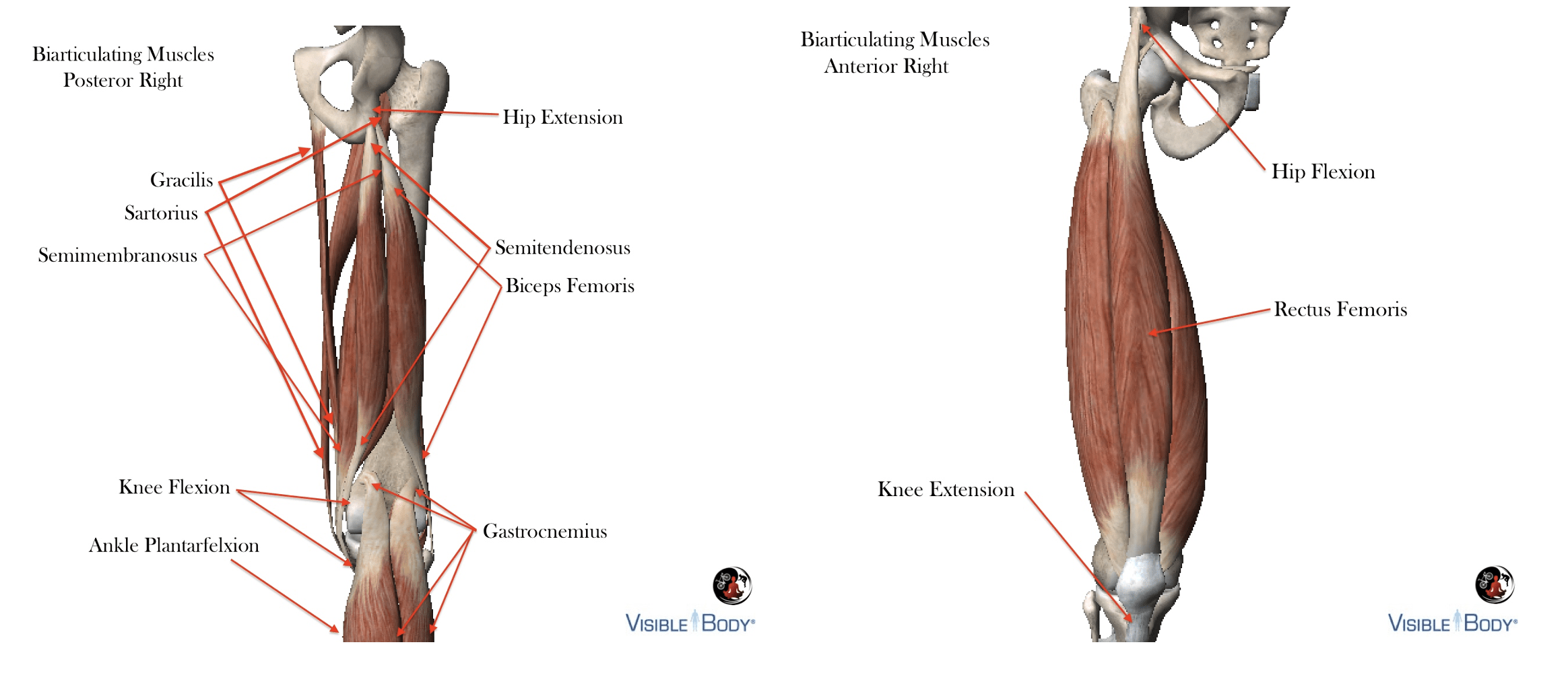
The biarticulating hamstrings extend the hip during the power-phase when pedaling, pulling ischial tuberosity and tibia (origin/insertion) towards each-other in concentric contraction. It is not possible for these two-joint muscles ot act exclusively at one-joint during extension (pedaling), so that the quadricpes, accompanying knee flexor moments as a byproduct of this shortening. Knee extension occurs regardless, because the quadriceps moments at the knee are superior during the power-phase when pedaling.
Research suggests that this phenomenon is a variant of Lombard’s Paradox – the paradoxical recruitment patterns of antagonist muscles that occur when moving from a seated position to standing.
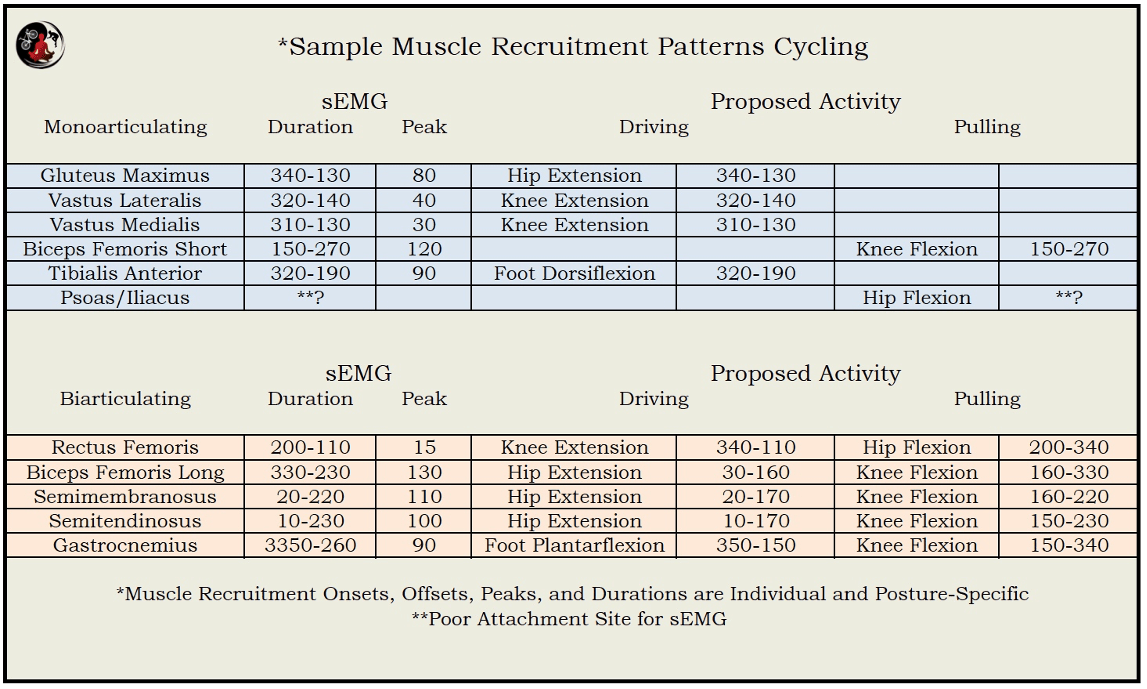
I conducted a small study (n=23/Road Cyclists) in 2013 to test contemporary research on muscle recruitment onset, offset and duration when cycling. The most important finding supported my notion that mean RMS values vary significantly between subjects and positioning – so be weary of muscle recruitment diagrams published online;).
Coactivation patterns were similar to other findings
How does position impact muscle recruitment patterns when pedaling?
Assuming that the pelvic angle is appropriate for an individual, the distribution of sagittal plane/anterior and posterior muscle recruitment patterns is determined by saddle and handlebar locations relative to the bottom bracket, and cleat position along the x-axis. If the variables that include saddle height, reach and differential are adjusted to maintain a constant hip angle, a more “aft” position relative to the bottom bracket tends to increase posterior tension/recruitment and vice-versa.
Increased posterior recruitment of biarticulating muscles can alter tibia on femur rotation in the direction of the biceps femoris (lateral) or pes anserinus (medial) as a function of morphology, pedaling base and resting muscle length.
In the same fashion pedaling base and cleat rotation impact coronal and transverse plane muscle function. Typically, exceeding the optimal pedaling base laterally increases medial activity/tension with greater lateral recruitment when the pedaling base is too narrow. Improper transverse plane lateral cleat rotation (medial foot rotation) increases tension at the biceps femoris and lateral rotators, with the muscles attaching to the pes anserinus and medial rotators impacted when a foot is forced into lateral rotation (internal cleat rotation).
Hopefully this entry helps readers appreciate the complexity and multivariate attributes of knee biomechanics when cycling.